The Eye
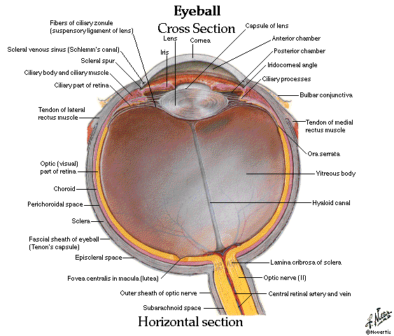
Basic anatomy of the eye:

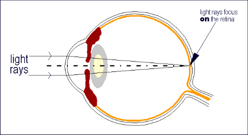
The eye as an optical system works like a camera. The information from the images we see, arrives at our eye with light rays. These rays encounter firstly the transparent cornea, then the lens and at last they focus on our retina. In short, the retina corresponds to the film of a camera.
The sharpness of our vision depends on the distance between the cornea and the retina as well as on the shape of cornea and lens. An eye without refractive errors is called emmetropic and the image is focused on the retina. If the distance between cornea and retina is incorect or the cornea does not have the appropriate shape, the image is displayed in front or behind the retina and vision is blurred. This situation is called ammetropia.

The main ocular refractive errors are:
All refractive errors can be corrected with the use of glasses or contact lenses -soft or rigid. A permanent correction with verygood results can be achieved by applying methods of refractive surgery. With the help of correction lenses the light rays change direction and focus onto the retina.
Conversely, in refractive surgery the curvature of the surface of the eye or the optical properties of the lens change and this results in vision improvement. The ideal refractive surgery candidate has stabilized refractive error for at least a year and an eye without any other problems. Contact lenses should be replaced with glasses for a short period before surgery.
A complete preoperative control of the eyes is absolutely necessary to personalize treatment. It takes two to three hours and includes measurments and eye examinations with special equipment. All results are recorded and used to choose the most suitable surgical procedure for the patient. The operation takes a few minutes under local anaesthesia and is painless.
.jpg)


